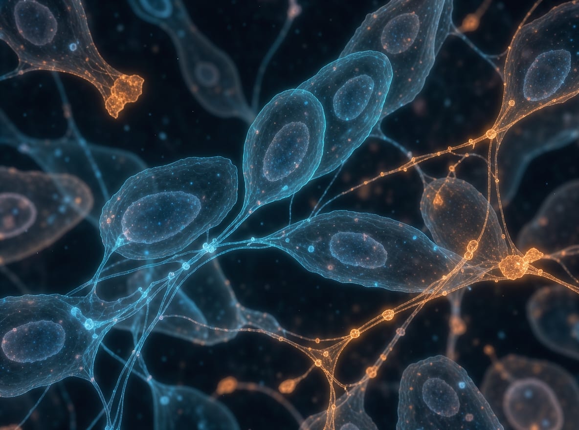
Cells sit in materials with different mechanical properties. A soft gel and a rigid surface may give a cell different resistance when it pulls on them. A fluid flowing across a cell can exert force along its surface. Mechanobiology brings biology, engineering, chemistry and physics together to study how forces and the mechanical properties of living material relate to cell behavior and tissue development [1]. Stiffness describes resistance to deformation. A force describes a push or pull. A cell can exert a pull on its surroundings, and the stiffness of those surroundings shapes how much they yield [2] [1].
Mechanotransduction is the conversion of a mechanical cue into an electrical or chemical signal [2]. The cue might be tension, pressure or the drag of flowing liquid, called shear stress [2]. A 2006 review of mechanotransduction across organs, tissues and cells described how the architecture of those systems can concentrate forces at sensitive structures such as membrane channels [8]. Starting at a cell's surface makes it easier to trace how a physical cue can reach different parts of a cell.
The matrix and the structures that connect to it
The extracellular matrix, or ECM, is a network of molecules outside cells. Collagen and other fibrous proteins help give the network structure. Some tissues also contain minerals in the matrix. A basement membrane is a thin sheet of matrix that underlies layers of cells. In other spaces between cells, fibers and water-rich material help the matrix bear compression [9]. The composition and arrangement of a matrix help define the surface or surrounding material a cell encounters.
Integrins are receptor proteins that span the cell membrane. The portion outside the cell binds to particular molecules in the matrix, such as fibronectin or collagen. On the inside, integrins connect through other proteins to the cell's supporting fibers [10] [3]. A focal adhesion is a grouping of integrins and associated proteins at such a connection. These groups often sit where actin fibers end at the membrane and can transmit both force and signals [3]. That location puts them in a position to carry a pull from the matrix inward and a pull from the cell outward.
A 2009 review of focal adhesions in work with artificial cellular environments discussed how the assembly of these groups interacts with forces arising from the growth of actin filaments and from contraction inside the cell [11]. The same 2009 review described integrin-based contacts responding to features of those surroundings such as how easily a material bends and how far apart binding sites are [11]. Imagine comparing the shapes of cells on surfaces that vary in stiffness while keeping the spacing of attachment sites fixed. Another comparison could vary that spacing while holding stiffness constant.
From the cell surface to the nucleus
The cytoskeleton is the dynamic network of protein filaments inside a cell. It helps give the cell shape and bear forces, and its filaments can assemble and come apart [12]. The actin part of the network can work with myosin motors to produce tension. A 2014 review of soft connective tissues at steady state described cells assessing the mechanical properties of the matrix through integrins and the actin and myosin network, then depositing, rearranging or removing material as part of maintaining the tissue [13].
The cytoskeleton also provides a physical path for force to reach the nucleus. Proteins at the nuclear envelope link the nuclear structure to cytoskeletal fibers [1]. Deformation of the nuclear lamina, a supporting layer at the nucleus, can influence gene expression [2]. Gene expression here refers to the use of genetic instructions by the cell. The line diagram follows a connection from the matrix through an integrin and the cytoskeleton to the nucleus. Chemical signaling and ion flow provide additional ways for a force cue to affect the cell [2].
A 2006 review of force transmission across tissues and cells described tension already present within those linked structures as a factor that can let several molecular responses take place together [8]. That concept helps distinguish the force a structure already bears from an added force. In a test of mechanical sensing, the starting mechanical condition is part of the description of the stimulus.
Piezo channels and the movement of ions
An ion channel provides a passage through the cell membrane for charged particles. Piezo1 and Piezo2 are mechanical sensors that allow positively charged ions, called cations, to cross when the channels open. Tension in a membrane can activate Piezo1. Piezo2 can produce an electrical response as cations move through its pore and change the voltage across the membrane [4] [5]. In this route the immediate signal is the movement of ions. At an adhesion, the signal can start with a change in force transmitted through connected proteins [2] [3].
In a 2010 study, experiments that reduced Piezo1 in a mouse neuroblastoma cell line established that the protein was required for the mechanically activated current measured in those cells [14]. In the same 2010 work, increasing the expression of mouse Piezo1 or Piezo2 induced currents with different timing, and reducing Piezo2 in dorsal root ganglion neurons decreased a rapidly adapting current [14]. The 2010 study proposed that Piezo proteins were components of mechanically activated cation channels based on those recorded currents [14]. Structural descriptions of the channels put the ion passage at the center of a complex that spans the membrane [4] [5].
Stiffness, gene regulation and cell fate
YAP and TAZ are transcription coregulators, meaning that they work with partner proteins to influence which genes get transcribed. YAP is also called YAP1. TAZ is the name used here for the coregulator also known as WWTR1 [6] [7]. Differentiation describes a cell developing the features of a more specialized type. Changes in gene expression help establish those features [15].
A 2011 study examining matrix stiffness and cell shape identified YAP and TAZ as relays of those mechanical cues to the nucleus, and reported that both the activity of the signaling protein Rho GTPase and tension in the actin and myosin network were needed for regulation in its experiments [16]. In the 2011 experiments, YAP and TAZ were required for stiffness-driven differentiation of mesenchymal stem cells and for the cell-shape-dependent survival response of the endothelial cells examined [16]. The survival measurement tracked whether the cells remained viable. Differentiation instead concerns the specialized characteristics a cell takes on [15].
A 2017 review of YAP and TAZ in cell and tissue research described how cues including shear stress, cell shape and matrix stiffness can be relayed into patterns of gene expression specific to a cell type [17]. As a review, it brings several settings together. A reader comparing experiments can ask which cue was varied, how a response was measured and which cell type was tested. Those details set the scope of a study's claim.
Gels give researchers a way to vary the stiffness of a surface while studying cells attached to it. A 2005 overview of quantitative research on tissue cells covered studies of cells adhering to gels or to other cells in which elasticity could be varied, and identified adhesions and the contractile actin and myosin network as important parts of the response [18]. Elasticity describes how a material deforms and returns toward its previous shape. The material under the cells in an experiment is called the substrate.
A 2006 study of mesenchymal stem cells in culture reported that soft, brain-like matrices favored nerve-related cell traits, firmer muscle-like matrices favored muscle-related traits, and more rigid bone-like matrices favored bone-related traits [19]. In the same 2006 culture experiments, blocking the action of nonmuscle myosin prevented the stiffness-dependent differences in lineage [19]. The measured result concerns the direction of specialization in a culture model with controlled matrix properties. The names of the tissues describe the elasticity being modeled and the traits compared.
Mechanical load in bone and tendon research
Wolff’s law describes the adaptation of bone structure to mechanical loads [20]. Research on bone asks how forces reach cells within its mineral-rich matrix and how those cells coordinate changes to the tissue [9] [20]. A 2006 review of bone remodeling described how bone-forming osteoblasts and bone-resorbing osteoclasts act in temporary groups that renew tissue, and discussed loading among the mechanical and molecular influences on turnover [21]. The mineral and fiber network bears loads, while the cellular process changes that network over time.
One way load can reach cells in bone is through the motion of fluid. Compression moves fluid through the matrix, where bone cells called osteocytes can sense its movement. Osteocytes can send signals to other cells involved in remodeling [20]. Distinguishing the site that senses the force from the cells that act in response helps to explain why bone mechanobiology spans more than one cell type.
A 2006 review of tendon mechanobiology described mechanical adaptation as changes in tendon structure, composition and material properties, and identified force sensing by tendon cells as an area where mechanisms remained incompletely understood [22]. A 2012 review spanning tendon development, maintenance and healing described changes in expression of matrix proteins and signaling molecules across the mechanical settings considered [23]. The reviews address different levels of observation. An account of material properties describes how a tissue bears force. A measurement of gene expression concerns the activity of cells in the tissue.
Mechanobiology in undergraduate research
The field's questions can appear in a cell biology lab that studies adhesion or gene expression, in bioengineering work that uses culture models, or in a materials lab interested in the surfaces cells encounter [1]. A useful starting point is to identify the mechanical feature on one side of an experiment and the measured cell response on the other. In an adhesion experiment that could mean a difference in the spacing of binding sites and a comparison of cell shapes. In a channel experiment it could mean a mechanical stimulus and a recording of current.
A project can also start with the material itself. For instance, a gel made to support cells has a stiffness and a surface where cells can attach. Measuring the gel's properties helps place later observations of cells in context. A description of the material, the cells and the comparison makes an experimental question easier to discuss at a first meeting.
The guide to finding an undergraduate research position covers how to start looking for a group, and the faculty conversation guide offers ways to talk about what a project studies and what a student could learn to measure. The glossary explains terms that appear across these papers.